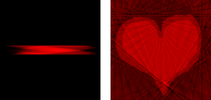
Happy Valentine’s Day! I might not be very romantic, but I had to do something with it. Imaging the heart is an important application of tomography and other techniques in medical imaging. Since hearts are everywhere on Valentine’s Day, let’s scan one. This article builds on my series of articles on tomography.
The image on the right, the sinogram, is the result of scanning the drawing of the heart in the same way that a medical CT (or CAT) scanner scans a person. See the mentioned series of articles for details. Now that I have your attention (because of the heart and it being Valentine’s Day when I post this), I want to seize the opportunity to push some science your way.
 Heart (left) and sinogram with 180 projections (right)
Reconstruction
After scanning, an image is reconstructed. This is the whole point of taking a CT scan, since you don’t have an “original image” for a real human heart, of course. In the image below, you can see that the reconstructed heart is quite close to the original one, but that they are not completely the same (it’s a bit blurred, and there is a haze in the corners).
 Reconstruction from 180 projections
The reconstruction becomes a lot worse if the number of projections is diminished, which is one way of lowering the X-ray dose of a medical scan. Below is a sinogram with only 18 projections, together with a reconstruction that was created from that sinogram. It is clear that 18 projections are not sufficient for a high quality reconstruction.
 Sinogram with 18 projections (left) and reconstruction (right)
Current Research
As an example of current research on tomography, the image below shows the result of a reconstruction with the DART algorithm that was developed at the Vision Lab of the University of Antwerp. For this reconstruction, the exact same projections were used as for the classical reconstruction that is shown above.
 Sinogram with 18 projections (left) and DART reconstruction (right)
This result is clearly much better. The DART algorithm assumes that the original image consists of homogeneous regions, and that the density of those regions is known. It then exploits this knowledge to create better reconstructions than the “classical” methods, with the same number of projections. Note that this is not directly applicable to medical scans, since people do not consist of homogeneous regions. However, it is directly applicable to things like materials science and non-destructive testing.
Add new comment